FREE STANDARD SHIPPING WORLDWIDE!
Research Papers
Sections 1 & 2
Recent research publications
Joint Pressure, Volume and Alignment in Development of AOA: Indications for Orthobiologics and Surgeons.
Published Date: 12-20-2019 Journal of Regenerative Biology and Medicine.
CONSIDERATIONS FOR THE DESIGN OF THE ‘IDEAL’ ANKLE ORTHOSIS
(Abstract version)
Section 3
Current research projects
Section 4
Future research (contact us for further information on our current and future research opportunities and directions)
There are thousands of ankle braces and many tape methods used both as safety devices and as medical equipment, on children and in the 'Workplace' that is Professional Sport in 2020. Traditionally the degree of protective ability is assessed by how much restriction of the STJ inversion or ankle roll they create, with greatest restriction deemed most protective, and by prospective studies in sports, comparing injury rates between methods. Whilst I contend that I could show wearing 'shoe boxes' on your feet instead of braces or tape ALSO reduces ankle sprains, for familiarity and comparison, we will be undertaking versions of the 'Shoe Box Study', but consider more of the consequences of the interventions.
Most Ankle surgeons do not expect much (and neither should you!) tissue repair/regrowth after lateral ligament injury, at best hoping that immobilization and Physiotherapy will lead to scar tissue. While some of the study directions may be unfamiliar and challenging, it is Ankle Surgeons and Doctors (who can prescribe and need evidence of ‘medical intent’ that makes sense), Athletes and often their Parents, for whom most studies are designed, as well as to develop initial proofs of new concepts for future funding. Therefore, the purposes of the of the studies are to demonstrate KiSS is superior to existing methods in;
Safety - We will use mechanical testing like for airbags, seatbelts or helmets, to measure the resistance of the KiSS system throughout normal inversion ROM and time, and Clinical prospective trials in sports v current methods. KiSS will be shown through physics and engineering to have sufficient energy storage capacity to 100% prevent a common ankle sprain whilst prospective studies in field and court sports will make direct comparison to existing common practice of tape and brace.
Repair of ligaments – We will examine the degree of ligament repair and joint integrity of an acute injury before and after KiSS intervention, where we propose to represent the world’s first non-surgical repair/regrowth of the lateral ligaments using the Isolated 3D method developed. No current system has ever been shown to repair ligaments, so follow up scans of ligaments following intervention has rarely been attempted nor has anyone attempted to isolate an image of them. Since KiSS contends to improve the integrity of the injured ligaments post intervention, so also requires a protocol to visually represent same. The initial Pilots are to develop methods and demonstrate the KiSS brace will improve immediate clinical outcomes in an acute injury phase and improve ligament integrity at 3 months post injury.
Early Mobilization – We will investigate what is known anecdotally, that when KiSS is applied to recent or ‘acute’ lateral ankle sprains, users clinically assess better regarding balance, mobility, pain, weight distribution, gait, comfort and perceived levels of support. The first step is to take one recent acute ankle sprain through KiSS rehab protocols, starting with early mobilization in the acute stage, compared to existing methods, whilst also taking before and after medical ‘Images’ of the ligament and joint integrity, utilizing the 3D isolation Technique.
Repair - As repair of the lateral ligaments is the ‘Holy Grail’ of ankle rehabilitation, it is our goal to produce supporting evidence for KiSS early intervention in acute sprains. These studies can and will be expanded to include comparisons across groups of alternative methods for comparison of medical benefit. Each new KiSS product will be subjected to mechanical testing for safety and shown to mobilize and improve outcomes for damaged or repaired ankle ligaments.
Regeneration/Orthobiologics- The clinical trials protocol will include the utilization of Orthobiologics, currently ineffective in ankles, but not in other joints where load can be induced internally and controlled. Orthobiologics (Stem Cells) act to enhance the growth of tissue and only KiSS can provide the joint support (Kinematic intra-articular Support) and ‘optimal loading’ necessary to regrow ligaments, so we have developed protocols to decide if KiSS truly is the world’s first Stem Cell or Orthobiologic Ankle system, with implications for prevention and repair of Ankle OA.
Section 1
Research and Publications
Lower Extremity Review Magazine
Editorial March 2023
What’s In A Name? That Which We Call Workout Shoes, By Any Other Name Could Be Personal Protection Equipment… Could It Not?
By Janice T. Radak, Editor
..."What Hubbard was really arguing for is a mindset shift. Moving away from the notion of ankle sprain as a 1-off injury that just needs a band-aid. Just as we wouldn’t ask someone with a partial foot amputation to simply stuff a sock in an old shoe, should we really ask athletes—or anyone who is being physically active regularly—to use a 50-year-old solution that current evidence doesn’t truly support?"
Lower Extremity Review Magazine
Editorial Aug ust 2020
What If We Adopted PPE as a Mindset for Ankle Protection?
By Craig J. Hubbard, Inventor/DesignerWorkplace safety, the right to be safe in your workplace and while going about the activities of your work, was certainly a thing before the COVID-19 pandemic struck. However, the pandemic laid bare some of the realities of the workplace that so many of us never even thought about. I’m talking, of course, about Personal Protective Equipment, or PPE...
https://lermagazine.com/issues/august/what-if-we-adopted-ppe-as-a-mindset-for-ankle-protection
Joint Pressure, Volume and Alignment in De velopment of AOA: Indications for Orthobiologics and Surgeons DOI: JRBM-1-011. Published Date: 12-20-2019 Craig J Hubbard
Founder- R&D, KiSS Ankle System Inventor/Designer - Biomechanics - KiSS Ankle Co, Australia Copyright© 2019 by Hubbard CJ. All rights reserved. This is an open access article distributed under the terms of the Creative Commons Attribution License, which permits unrestricted use, distribution, and reproduction in any medium, provided the original author and source are credited.
Abstract
To fix and prevent, not ‘manage’ Osteoarthritis OA in the lower limb, should be the collective ‘Holy Grail’ with or without Orthobiologics OB. Lateral Ankle Sprain LAS, Chronic Ankle Instability CAI and Ankle Osteoarthritis AOA create asymmetry which alters the biomechanics of the entire lower limb, so by better addressing AOA, we can probably do more than just impact the multi-billion dollar annual costs of AOA.
It would seem we have advanced, if not futuristic Surgical techniques and Orthobiologic technology, so what is missing? The short answer is Medical Intent MI. Devices and methods used in rehabilitation need MI intent to enable and stimulate repair. The world is changing and Morals Ethics and the human costs, are being counted. CTE concussion is just the tip of the iceberg for cumulative trauma injuries, in cost and prevalence, and class actions seek to defend and enforce people’s rights to safety, either in the workplace, as in professional sport, or in medical outcomes.
The significant yet hidden role of the subtalar joint as a ‘Safety Valve’ was first noted by Albert Ferguson in 1972, yet today the contradictions in rehabilitation and injury prevention devices that restrict the STJ, remain commonplace. It is also necessary to consider what has changed inside the synovial capsule before and after lateral ligament injury, such as pressure, joint alignment and space, so we can better understand and design for restoration of homeostasis.
This paper will examine factors, causes and interventions that may be inadvertently restricting or preventing Orthobiologics effectiveness in the human ankle from a Translational Medicine perspective and an engineering bias.
You can view the article here.
Section 2
CONSIDERATIONS FOR THE DESIGN OF THE ‘IDEAL’ ANKLE ORTHOSIS
(ABSTRACT VERSION)
BY
Craig J. Hubbard, B. App.Sc.
Contact: [email protected]
Australia
ACKNOWLEDGMENTS:
The Author wishes to thank the following people for the assistance in preparation of this paper. Dr R Borrenger and Mr Ron Avery, Cumberland College of Health Science, for their co-operation in providing access to anatomy “wet” laboratory. Mr George Gedge, for his encouragement of the original idea for this study. Mr Andrew Cogian, for his help in constructing the surveys. Those athletes and related professional who took the time to complete the surveys.
Contents
Anatomy and functional anatomy
Ankle and subtalar joint motion and stability
Lateral ankle sprain mechanics
Functional instability
Taping and Bracing as ankle injury prophylaxes
Implications and contraindications
Design Considerations
Anatomy and functional anatomy
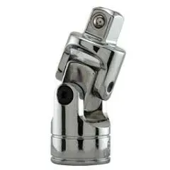
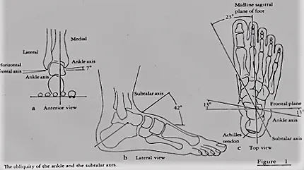
According to Wright (32) the ankle and subtalar joints function similar to a universal joint. When motion parallel to one plane is limited, for example the ankle axis during rotation of the lower leg, motion must occur at the other joint, in this case rotation about the subtalar axis. Reciprocally, where STJ ROM is restricted or exceeded in supination, the analogy of the universal joint means that this motion must then be resolved at the ankle joint, through the restricted motion of internal rotation of the talus within the ankle mortise; a motion limited by the lateral ligaments. With loss of subtalar motion (for any reason), the ankle has no relief from superimposed rotational forces as the leg rotates (43).
During a lateral ankle sprain the anterior talofibular ligament ruptures first as the limit of STJ ROM is reached, allowing the fibula to slide posteriorly, releasing the leg to externally rotate. As Rotation progresses, the calcaneofibular ligament is stressed and is the next to rupture. As this happens the loading appears to shift to the medial dorsal talus. This observation is consistent with the findings of Bruns and Rosenback (40) who demonstrated pressure increases on the medial talar border at a similar stage of ligament dissection. Along with other researchers (41,42,43) they have related the incidence of posteromedial osteochondral lesions to a history of lateral ankle sprains. Glick et al (24) however have demonstrated radiographically the inability of rigid tape to hold the talus within the ankle mortis for any time longer than 20 minutes and was in fact probably effective for less. Larsen (55) after similar findings had good reason to doubt the validity of ankle taping as a prophylaxis for chronically unstable ankles.
Figure 1 The obliquity of the ankle and the subtalar axes
(a) Anterior view(b) Lateral view (c) Top view
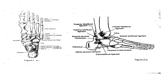
The shape of the articular surfaces is particularly important to the evaluation of components of joint motion. Ligaments guide and check excessive joint motion with fibre direction determining what motions are guided and limited (33). The ligaments which stabilise the ankle consist of the strong medial ligament, and the 3 bands of the lateral ligament. Together with the lateral malleolus they provide lateral stability to the ankle joint and stabilise the talus within the ankle mortise (34) (figure 1b and 2 b).
Figure 2
Ankle and subtalar joint motion and stability
During contact the talus and lower leg function as one unit, the calcaneus and foot as another. During this phase the foot is fixed to the surface so that
any rotation of the leg must be resolved through rotation at the STJ (32).
Lateral ankle sprain mechanics
Rotary stability in a horizontal plane is provided by tension in the collateral ligaments and by compression of the articular surfaces. It is suggested rotary instability may be an additional factor in patients whose symptoms persist after injury to the lateral ligaments (35). The ankle and subtalar axes are shown in figure 3.
Figure 3
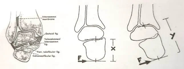
The AJ rather than the STJ is likely to be damaged during inversion stress since the thick and strong inter osseous talocalcaneal ligament (internal) and the joint capsule which maintain the joint’s integrity are both close to the axis and to the applied force (figure 5a). When an inverting force is applied to the calcaneus a rotary torque is created about the STJ axis (figure 5b) along the moment arm X. When the limit of STJ motion is reached, the calcaneus and talus would then become a rigid lever Y (figure 5c). The torque is then transmitted to the ankle joint along this longer moment arm Y. If the loading force is rapid and is not resisted, the high impulse moment tending to separate the joint laterally may be greater than that which was applied about the subtalar axis.
Functional Instability
Functional instability (FI) of the ankle refers to repeated sprains or giving way of the ankle (freeman et al 1965) cited by (8, 19, 20, 23) and was a residual disability presenting in 20-40% of inversion sprains (19, 21). Smith and Reischl (46) report residual symptoms in 50% of young basketball players following lateral ligament injury. It has been proposed that injury my lead to a partial deafferentation of peripheral reflex mechanisms (23). Konradsen and Raven (23) measured the peripheral and central reaction times in unstable and stable subjects using a tilting trapdoor (figure 4a).
Figure 4 (a)Ankle inverting platform (45)page 73 (b) Postural adjustment tosudden ankle inversion (23) page 389.
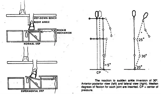
They were able to show that peripheral reaction time was increased significantly (p <0.01) in FI subjects (84 ms vs. 69 ms) and that central reaction time was uncharged (20 ms). Central reaction time was the time from the first peroneal response to the first hamstring or quadriceps response which indicated supraspinal postural adjustments and a pressure relief through a shift in the center of pressure (figure 4b) (23). Springings and Pelton (45) demonstrated that the time for the foot to invert through 30 degrees stepping down from a height of 30 cm onto a collapsible platform was in the vicinity of 150 ms. It is postulated therefor that functional mechanisms will be insufficient to prevent lateral ligament injury during most sporting activities.
From this discussion it can be seen that during even mild inversion stress, motion could well progress past the midpoint of motion before any spinal level reflex can be anticipated (Peroneus Longus 65 ms, Peroneus Brevis ms), and still a further 20 ms is required before postural adjustments which further absorb shock and relieve pressure, can take place (23) (see figures 4a, 4b). Since it is anticipated that most sporting landings would be more rapid than the step down studied,
inversion stress could easily disable reflex as a preventive mechanism (52).
Taping and Bracing as ankle injury prophylaxes
The principle of ankle taping with its support structure established by anchors, stirrups and heel locks of rigid tape is widely used and recommended. The reports of the behaviour of this structure under exercise conditions were reviewed by the Author to identify the causes of loosening during exercise (62). The findings of this review are summarised.
The lateral stirrup and anchor sites were most affected, with tearing of the stirrups and/or displacement of the stirrup and anchor down the leg. Loosening occurred below each malleolus with the tape later functioning as a canvas boot.
Tensile stress concentrations within the stirrups, high impulse loading and shear stress at the skin – anchor and anchor – stirrup junctions were believed to be responsible for the loss of support strength as the foot attempted to accommodate a normal range of motion.
Perspiration contributed to loosening, affecting adhesion and inducing creep.
Statements of researchers such as Delacerda (18) that “… the purpose of ankle taping is to reduce joint range of movement….” (p 78) have reinforced the traditional belief that to protect the ankle from injury it is necessary to restrict the ankle from inverting by limiting the ankle range of motion (ROM) in this direction.
Garrick and Requa (2) have proposed a theoretical aim of ankle taping “…to support externally a ligamentous structure without limiting normal range of motion or function. This support of ligaments need be present only when the physiologic or normal ranges of motion have been exceeded.” (p 202). They also contend, “….the achievement of this goal, however, is virtually impossible.” (p202).
Further, “… one must establish that restriction of normal motion is an adequate indicator of the protective influence of the method of support and has no other deleterious effects.” (p202).
Gross, Lapp and Davis (17) evaluated the comparative effectiveness of Swede-o-Universal Ankle support (SO) (currently being used at the Australian Institute of Sport), Aircast Sport-Stirrup (AS) and ankle tape in restricting eversion-inversion ROM before and after exercise ( 10 minute figure eight running and 20 unilateral toe raises). The found that all support systems significantly reduced eversion and inversion both before and after exercise.
Robinson, Frederick and Cooper (27) used progressive stabilisation with rigid inserts insides high top basketball shoes and performance times on an obstacle course to examine the effects of systematic changes in ankle support on range of motion and performance. Their results showed that systematic changes in ankle and subtalar ROM did measurably and significantly affect performance. Several points raised in their discussion were of interest.
Firstly, “Directional changes require a large horizontal component of force. Positioning the leg while manoeuvring is accomplished by the normal ROM at the ankle and subtalar joints. If ankle motion is restricted, then the ability to position the leg to apply a large horizontal force component is reduced, decreasing manoeuvrability.” (p 627).
Secondly, in agreement with Garrick and Requa (2), that support need only be present at the extremes of range, Robinson et al (27) state, “Obtaining this goal with current technology and traditions is improbable, therefore, prophylactic ankle support becomes a question of balance, with protection and performance at opposite ends of scale.” (p 627). Lastly, they suggest,
“Further work is needed to examine the concept of an optimum ROM and restriction for adequate performance and protection.” (p 628). The comments of Garrick and Requa in 1973 that support at the limit of range of motion is “virtually impossible” (p202) can be understood in the context of materials and methods then available.
To suggest a compromise between natural function, performance and injury prevention, serves only to emphasise the degree of stagnation present within current ankle support design, research and use.
Implications and contraindications
The restriction of natural range and function, difficulty of application and comfort considerations are some of the major reasons why sportspersons frequently compete without external prophylaxes. Since ankle prophylaxes reduce inversion ROM it can be anticipated that ROM reduction will reduce the capacity for the motion and range dependent responses to inversion stress.
The limit of motion is reached more quickly when inversion ROM is rigidly restricted and possible more frequently. Protective actions of peroneal reflex and postural adjustments are proportionately less likely to have evolved effect. The contraindications of this are that normal and abnormal inversion stresses, instead of being dissipated by muscle and postural adjustments which spread the stress over time are transferred to the leg with an impulse dependent upon the rigidly of the support material.
The effects of these repeated low loadings and occasional high impulse loadings upon the development of over-use syndromes and the ligamentous integrity of the lower limb respectively, has not been investigated. With the development of quantitative assessment techniques, it should be possible to demonstrate the effects of limitation of natural range imposed by current techniques. It will not only require that it be shown that range restrictions are detrimental to performance, function and ligamentous integrity (if this is in fact so), but that an alternative must also be available. These concerns are consistent with Ferguson (1), and relate to the importance of the “ankle safety valve”.
Design Considerations
Improvements indicated at the skin- anchor junction are directed at enhancing shear holding, stress and perspiration dissipation, whilst more effective attachment of the stirrup to the anchor is implied.
By making the stirrups from an elastic material capable of storing and transmitting injurious forces over time to the anchor sites, the impulse related to stirrup and anchor displacement in taping is reduced. Injurious forces imposed on the calcaneus will be substantially dissipated during rotation about the subtalar joint with dissipation of this force to the lower limb through extrinsic (attachment to the skin) and intrinsic (muscle forces, postural adjustments) pathways.
It would be desired to make the smallest size available to young children so that they can be protected from an early injury which could present with future complications (functional instability, osteochondral lesions, etc.).
To ensure ease of application and diversity of use the substantially re-useable brace is implied. This is a consideration for use in rehabilitation since access to treatment modalities in desired.
An ankle orthosis should be able to provide minimal restriction of motion at the neutral position, with support increasing to maximum at the elastic limit of joint motion.
Adjustment is desired for selecting the required amount of inversion, plantar flexed inversion and for correction of talar alignment at heel strike. The features are essential if the orthosis is to be used to non-rigidly correct rearfoot varus and valgus conditions.
During rehabilitation active range of function must be controlled so that appositional healing of ligaments is encouraged. This is achieved by superiorly directed non-rigid support of the talus within the ankle mortis for extended periods.
The maintenance of ankle joint articular surface contact must be achieved however without causing compensatory motion of the talus in the ankle mortis as a result of restriction of subtalar range of motion.

Section 3
Current Pilots & Research
KiSS Ankle System - University of Sydney Faculty of Health Sciences collaboration
This document describes the collaboration between the Musculoskeletal Health Research laboratory at the University of Sydney and KiSS ankle brace.
University of Sydney Musculoskeletal Health Research Lab: The MSKH group at the Faculty of Health Sciences has a vision to cure pain and disability associated with musculoskeletal conditions. Our mission is to conduct excellent well-funded collaborative multidisciplinary research which is highly translational to prevent, treat and reduce the burden of pain and disability associated with musculoskeletal conditions. We are interested in assessing the mechanistic and clinical effects of the KiSS brace. Trial aims will be decided by collaborative agreement, but the study design, method, analysis, and results will be conducted independently from KiSS.
KiSS Ankle Systems are designed to provide two substantive benefits over existing braces and taping methods. Those benefits are superior ‘safety’ when used for prevention and ‘medical intent’ to provide ‘optimal loading’ of ligament and cartilage for ‘innate’ or Biologically and/or surgically assisted repair/regeneration. To date no method or device can reduce sprains without considerable compromise of function or natural responses to injury. Further no device has ever been shown to maintain the talocrural joint over time, hence neither mobilize acute sprains nor repair lateral ankle ligaments.
Project 1
We propose a crossover study of about 30 participants with CAI. After baseline measurements, half of the participants will start using the brace while the other half will continue without the brace. At 3 weeks the groups will swap interventions. Clinical and biomechanical tests will be done at baseline, pre and post brace application), 3 weeks and 6 weeks. 16 participants (8 in each group) will also undergo a biomechanical evaluation.
Equipment: 30 braces of different sizes, 16 camera/6 force plate biomechanical lab, SEBT platform, isokinetic dynamometer, Zeno, tape measure, goniometers, stop watch, Smartspeed equipment.
Biomechanical experiments will be performed on 16 healthy young adults. Data will be recorded in the Biomechanics laboratory at the University of Sydney after ethics approval from the University’s Human Research Ethics committee. Each participant will provide written informed consent prior to participating in the study. All participations are to be able to walk unassisted and free from any pathology that could influence their ability to walk normally.
Each participant will perform 3/4 sports tasks while wearing and not wearing KiSS; these sports tasks are side-cutting, vertical jump, drop-landing and drop-jumping. All participants must be familiar with performing these tasks and will be given appropriate familiarisation time. Kinematics and force-plate data will be recorded simultaneously for each trial. The three-dimensional positions of the retro-reflective markers will be attached to each subject and recorded using a video motion capture system (Motional Analysis Corporation) with 16 cameras sampling at 100Hz. Ground reaction forces are to be measured using 5 piezoelectric charged force plates (Kistler Corporation) with a sampling rate of 100Hz.
Scaled-generic linked segment models will be developed by scaling a 10-segment, 23 degree-of-freedom generic model of the body. Scaled-generic models will be created by scaling segment lengths and segment inertial properties for individual subject anthropometry. For each instant of the task duration, a set of optimal joint angles will be calculated by minimizing the sum of the squares of the differences between the positions of markers defined virtually in the computer model and the reflective markers adhered to the subject. The net joint moments will then be computed by applying the calculated values of joint angles and measured ground reaction forces to the model using inverse dynamics. Results will then be averaged across all subjects for each condition (with and without KiSS). One-way repeated measures ANOVA will be used to detect statistical significance for each dependent variable between the KiSS- and no- intervention groups.
We ship high quality Hand-made Australian Ankle systems worldwide for Athletes, Surgeons and Weekend Warriors
Decades waiting for Social Media, Science & Sports Litigation to expose the harm so we can bring you the safest ankle system on the planet.
We have developed & implemented our own 'Crash' & Mechanical Tests, using the product designer as a 'Crash Test Dummy.'
KiSS Ankle Co
Laurieton NSW Australia
Tel: International +61497170013
In Aus: 0497170013
Or email the Boss: [email protected]
Website By Abnormal Marketing
KiSS Ankle Co ABN 44576885484
